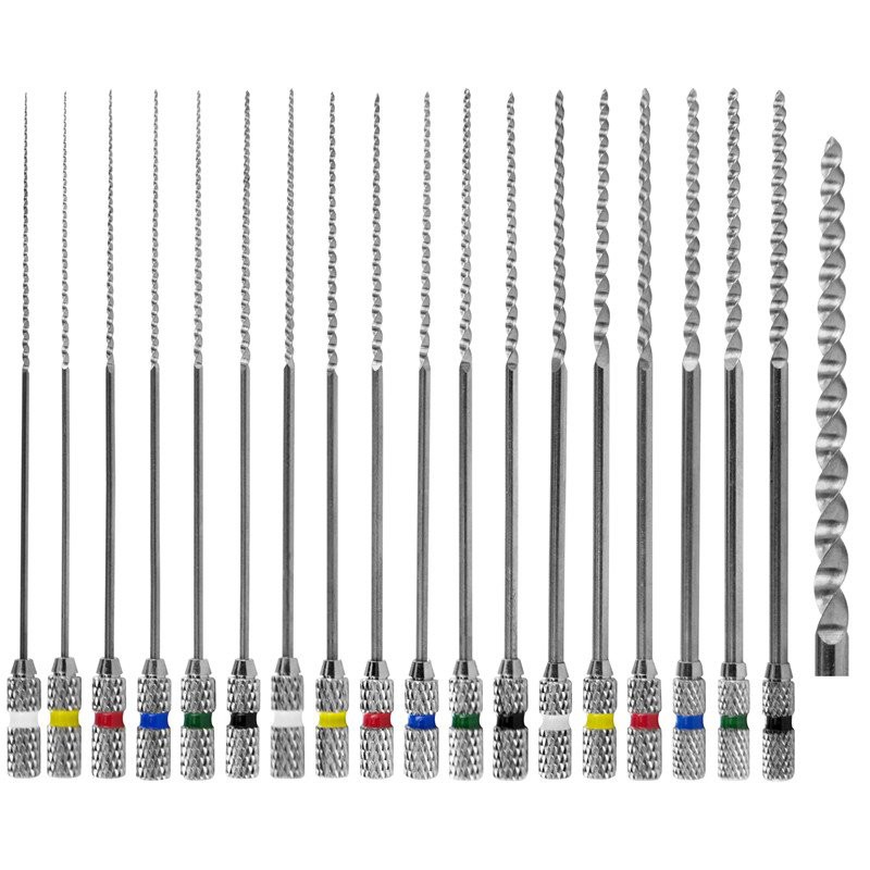
The main purpose of root canal treatment is to eradicate the infected microorganisms in the pulp, and eradicate the inflammatory reaction caused by the periapical tissue by disinfecting and sealing the root canal space. Successful pulp treatment depends on proper mechanical shaping and sufficient effective irrigation plan. As well as the three-dimensional filling of the root canal system, a long-term treatment success was finally obtained. However, complex changes in the root canal system usually affect the effect of root canal treatment and even lead to treatment failure. Therefore, emphasizing the diversity of root canal morphology and being familiar with the anatomical morphology of the root canal is of great significance for improving the success rate of clinical treatment.
Generally, we think that the configuration of the root canal system of the anterior teeth, especially the maxillary central incisor, has a simpler change than the posterior teeth. Altman et al. classified the anatomical morphology of 100 maxillary central incisors and studied the changes in their roots and root canals. They found that almost 100% of the teeth have only one root canal, and the special root canal morphology of the maxillary central incisor exists. The probability is very low.
Therefore, the multiple canal structure and special root canal configuration of the maxillary central incisor are easily overlooked. If no excess root canals or remaining infected parts are found during the treatment, and they are cleaned and sealed, it may continue to stimulate the periapical tissue of the affected tooth, thereby affecting the prognosis of root canal treatment. In the process of root canal treatment, children’s teeth are usually unclosed apical foramen and are flared, and due to underdevelopment and relatively large pulp cavity, it is necessary to determine the working length and choose the root canal filling material for the root canal. More difficulties came. As early as 1984, Vertucci divided the root canal morphology into eight types according to the number and orientation of the root canals and the number of apical foramen, including type I. A single root canal extending from the pulp cavity to the apical foramen has a single root canal. Apical foramen; Type II, two independent root canals leave the pulp cavity and merge above the apex to form a single apical foramen; Type III, a single root canal leaves the pulp cavity and divides into two canals, and then merges into In a root canal, there is a apical hole. And there are IV type, two independent root canals with two independent apical foramina; V type, a single root canal leaves the pulp cavity, and then divided into two root canals, with two apical foram Two independent root canals leave the pulp cavity, merge into one, and then divide into two root canals with two independent apical holes.
The multiple root canal structure and V-shaped structure of children's maxillary central incisor are special root canal anatomical changes, and there are more procedures and precautions in root canal treatment. An eight-year-old child suffers from repeated swelling of the gums in the area of the front teeth and painful bite. Clinical and X-ray examinations in the oral cavity showed that the No. 21 tooth was broken at 1/3 of the neck, and there was no obvious loosening in the intraoral exploration and percussion. The apical X-ray film showed that the root canal was transparent, and the apical foramen showed an open state. The pulp cavity was relatively wide and was diagnosed as irreversible pulpitis, and a root canal treatment plan was made.
First, the periodontal area of the tooth is anti-inflammatory, antibiotics are used and the state of the periapical area is observed. After the root development is completed at the age of ten, the root canal treatment of the tooth is planned and the tooth defect is repaired. During subsequent follow-up, it was found that the shadow of the root tip of the tooth gradually became smaller, and X-rays showed that the root canal was translucent, the length of the root tip increased, and a root tip barrier was formed, ready for root canal treatment. The teeth were isolated with rubber dams, and root canals were explored with #10 and #15 K files, and then the root canals were prepared with a nickel-titanium machine file system, and 5.25% sodium hypochlorite and physiological saline were used for extensive irrigation. Subsequent X-ray photos taken from various angles showed that radiotransparency still exists in the root canal. Using cone beam computed tomography (CBCT) scans, it was found that the root canal was close to one-third of the root canal in the mesial direction. There are bifurcations and two independent apical holes, which are identified as the V-shaped structure of the maxillary central incisor. Continue to unclog the root canal under a microscope to determine the working length of the root canal again, and then prepare the root canal with a nickel-titanium machine file system. After cleaning the root canal with 5.25% sodium hypochlorite and normal saline, it is temporarily sealed with calcium hydroxide for two weeks. After that, the rubber dam was used to isolate the teeth again, the root canal was dried after flushing with 5.25% sodium hypochlorite and normal saline, and then the root canal was filled with gutta-percha and sealing compound, and the three-dimensional sealing of the root canal was achieved by the vertical compression technique of hot gutta-percha. Finally, use composite resin to restore the teeth. The patients were followed up three months, six months and twelve months after endodontic treatment. The clinical examination showed that the teeth were not painful under knocking, the gum tissue was normal, the periodontal detection depth was 2–3 mm, and there was no abnormal looseness. The apical X-ray film showed that the periapical period was normal, there was no external absorption, and the periapical period and the periodontal ligament were in a continuous state.


